MY FAVORITE TISSUE - THE AVIAN BEAK
Welcome!
Hello!
My name is Rachel Sorensen and I am a fourth year biology student. I’ve
designed this blog as an assignment for the BIOL 3500 (Histology) course at
Memorial University of Newfoundland. Hopefully my series of posts can help you
learn a little about the histology of bird beaks, and how the specialization
of these tissues act as a key characteristic for the success and continued
biodiversity of avian species.
Who are the Class Aves?
Class
Aves is a class of vertebrates that includes the birds. Although birds are a
close relative to the reptilians and live in close association with the
mammals, this division of animals has numerous specializations that distinguish
them from other animals across the globe. While characteristics such as
feathers, fused metatarsals, a unique digestive system, and the ability to
engage in flapping flight are important indicators of this class, the evolution
of a toothless beak is equally critical. The avian beak and its morphological
flexibility between orders, families, and species of birds has not only allowed
permitted birds to become specialized in diet relative to other animals, but
has granted birds the ability to diversify and colonize a variety of ecological
niches relative to one another (Bhullar et al., 2015).
.")
Origin of the Bird Beak: Way Back When...
The
evolution of avian biodiversity and specializations of the bird beak spiked
just after the end-Cretaceous extinction
event (Bhullar et al., 2015).
DID YOU KNOW?
Birds actually evolved
from REPTILES!
- Ancestral reptiles had paired, compact premaxillary bones, giving them a snout to explore the environment.
- Although this greatly contrasts what we visualize about birds, the premaxilla is also a very important bone in the internal anatomy of the beak. In modern birds, the premaxilla bones have become fused and elongate.
Figure 2: Comparison of reptilian and avian facial characteristics, demonstrating the fused and elongate premaxilla of birds.
The Big Picture
Although the fully developed forms of bird beaks are highly variable, the basics of gross
anatomy stay consistent amongst species (Van Hemert et al., 2012).
Deep
inside the bird beak is a framework of
bone, which is supplied by a
vascular layer of blood vessels and nerves. Externally, what covers these
layers and makes the beak so visually interesting is a thick layer of keratinized epidermis (Chewy Editorial, 2018).

The
keratin forms a sheath around the beak, otherwise defined as the rhamphotheca (Macwhirter, 2009). The
sheath enclosing the upper jaw is the rhinotheca,
while the sheath along the lower jaw is the gnathotheca.
Figure 3: Rhamphotheca covering the avian beak, divided into the upper rhinotheca and lower gnathotheca (Pesek, 2001).
INTERNAL ANATOMY - BONES

The beak consists of upper maxillary
bones and lower mandibular bones
(Chewy Editorial, 2018).
DID YOU KNOW?
To effectively adapt to
flight, birds have evolved pneumatic
bones in several areas, including the skull. This means there are pockets of air within the tissue, making them much lighter than mammals (Jacob, 2018).

INTERNAL ANATOMY - VASCULARIZED LAYER
While the external layer of the bird beak is
keratinized and the internal layer is composed of bone, there are important
structures for respiration and olfaction
that lie in between (Van Hemert et al., 2012).
The
nasal passages on the upper beak are lined with hyaline cartilage, surrounded by connective tissue, abundant in elastin
and collagen fibres. In terms of
epithelia, there are different categories of cells localized to different
locations of the beak.
- Stratified squamous
epithelium lining the rostral nasal chamber.
- Respiratory epithelium in the middle nasal chamber and infraorbital sinus. This is pseudostratified,
ciliated, columnar epithelia.
- Olfactory
epithelium in the caudal chamber. This involves a layer of simple
cuboidal cells.
On
both the upper and lower portions of the beak, there are also salivary glands. These are exocrine
glands, and assist in lubricating food entering the digestive system
(Widlife Rehabber, 2018).

Figure 5: The upper beak of a Black-capped Chickadee (Poecile atricapillus)(Van Hemert et al., 2012). A) Basement membrane, salivary glands, and cartilage of the rostral nasal passage. B) Hyaline cartilage, pseudo stratified respiratory epithelium bearing cilia, and mucus glands of the nasal passage. C) Salivary glands and simple cuboidal olfactory epithelium. D) Hyaline cartilage and stratified squamous epithelium of the rostral nasal passage.
 The
highly vascularized layer of the beak is the dermis, which is concentrated with blood vessels and nerves
(Van Hemert et al., 2012).
The
highly vascularized layer of the beak is the dermis, which is concentrated with blood vessels and nerves
(Van Hemert et al., 2012).
The dermis becomes progressively thicker nearing the
beak tip, and contains branches of trigeminal nerves, facial nerves, arteries,
and veins.
The
dermis also has papillae surrounding
the circumference of the tissue, and stopping just before reaching the
keratinized layer. The papillae function to bring nutrients and oxygen to other
layers epidermis.
There are also HERBST CORPUSCLES present within the dermis, in close association with
the bone tissue of the premaxilla and lower mandible (Van Hemert et al., 2012).
WHAT ARE THEY?
Herbst corpuscles are nerve endings that function as touch receptors, very similarly to the
well-known pacinian corpuscles (“Corpuscles of Herbst”, 2018).
WHY DO WE CARE?
"Wading birds” are well known for having high concentrations of herbst
corpuscles in the pits of the mandible.
- Wading birds are species located on
shorelines and mudflats, including cranes, herons, flamingos, and many others.
- Having a high concentration of these touch receptors in the beak allows the
birds to sense prey beneath damp substrate.


FUN FACT!
A
common coloration characteristic of bird species is a distinct black beak.
This trait is ALSO attributed to the
tissue inside the beak, which hosts an abundance of melanocytes (Van Hemert et al., 2012).
- Melanocytes are melanin-synthesizing cells, derived from the neural
crest cells in the stratum basal of the epidermis (Derm 101, 2018).
Figure 8: The Royal Spoonbill (Platalea regia), a New Zealand species showing a striking black beak.
EXTERNAL ANATOMY:
IT'S NOT ALWAYS ONLY THE INSIDE THAT COUNTS!
As you’ve already learned, the rhinotheca and gnathotheca are composed of the protein KERATIN,
sheathing the beak.
Specialized
epidermal cells, the keratinocytes, produce keratin as part of the keratinization process (Fusenig et al.,
1980). Keratin is defined as a fibrous
protein with molecules of sulphur, forming the basis of horny epidermal
tissue (“Keratin”, 2018).
SO HOW DOES THIS WORK?
 The
basement membrane separates the epidermis from the dermis (Van Hemert et al.,
2012). Proliferating epidermal cells are located at the stratum basale of the epidermis,
attached to the basement membrane (Gilaberte et al., 2016).
The
basement membrane separates the epidermis from the dermis (Van Hemert et al.,
2012). Proliferating epidermal cells are located at the stratum basale of the epidermis,
attached to the basement membrane (Gilaberte et al., 2016).
At
this stage, keratin is already primitively formed, but has not yet packed nor
formed cross-linked disulfide bonds to provide the strength observed in later
versions of the tissue.
Keratinization involves numerous maturation steps,
leading to cell death and degeneration of nuclei and organelles in the upper
layers of the epidermis.
Figure 9: Layers of the epidermis.

The dead cells become engorged with keratin, forming
the stratum corneum, which acts as a horny outer layer of the epidermis. Due to the action
of corneodesmosomes, the cells are strongly linked
together, providing an insoluble barrier to the surface of the beak
(Ishida-Yamamoto & Igawa, 2015).
Figure 10: Beak of a Black-capped Chickadee showing mild hyperkeratosis of the stratum corneum (Van Hemert et al., 2012).
 The mature keratinocytes conform to the general shape of the beak, to maintain smoothness and structural integrity.
However, the
keratinocytes making up the superficial layer lining the beak are not arranged
uniformly (Van Hemert et al., 2012). This surface may show signs of abrasion, due to
the occasional rambunctious activities in the life of a bird!
Figure 11: Two sassy sparrows - an example of how beak abrasions may be inflicted in the wild!
SO YOU KNOW ABOUT STRUCTURE, NOW WHAT ABOUT FUNCTION?
As you've learned,
birds have become extremely diverse based on the variability of beak shapes, which allowed them to occupy and succeed in different ecological niches.
Here are some COOL functional examples of the avian beak!
1. THE GREAT BLUE HERON (Ardea herodias)
This bird has a beak like a spear ("Great Blue Heron", 2018).
- Because the Great Blue Heron feeds primarily on small fish in an aquatic environment, this beak shape allows the heron to jab its prey.
 Figure 12: A Great Blue Heron using it's spear-like beak to feed on fish.
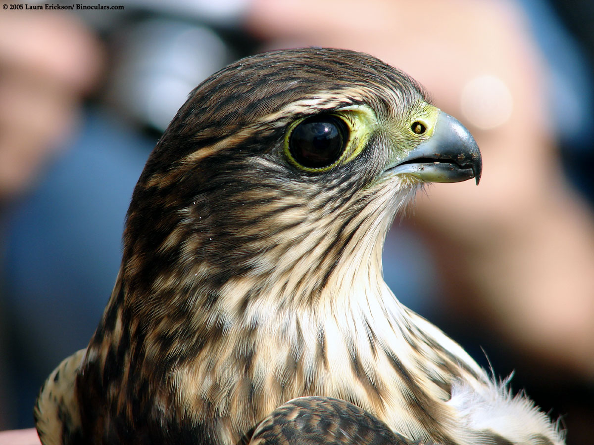
2. THE MERLIN (Falco columbarius)
The Merlin is a species of falcon. This bird has a sharp point on the tip of it's beak ("Merlin", 2018).
- Because the Merlin feeds primarily on other birds by catching them through a high-speed air chase, this sharp point allows this species to tear flesh apart.

Figure 13: Merlin exhibiting sharp point on tip of beak, used to function as an active predator (Photos by Laura Erickson, 2005, and Nancy McKown, 2014).
3. THE WHIMBREL (Numenius phaeopus)
This species has a long beak that curves downward ("Whimbrel", 2018).
- This allows the Whimbrel to dig, pulling crabs and worms out of the substrate.

Figure 14: The Whimbrel digging a crab out of the sand using it's long, down-pointed beak (Photo by Alan Vernon, 2007).
REFERENCES
Bhullar, B. S., Morris, Z.
S., Sefton, E. M., Tok, A., Tokita, M., Namkoong, B., . . . Abzhanov, A.
(2015). A molecular mechanism for the origin of a key evolutionary innovation,
the bird beak and palate, revealed by an integrative approach to major
transitions in vertebrate history. Evolution,
69(7), 1665-1677. doi:10.1111/evo.12684
Chewy Editorial. (2018).
Bird Beak Anatomy. Retrieved from https://www.chewy.com/petcentral/bird-beak-anatomy
Corpuscles of Herbst.
(n.d.). Retrieved from https://www.revolvy.com/page/Corpuscles-of-Herbst
Embryologic, Histologic,
and Anatomic Aspects. (2018). Retrieved from https://www.derm101.com/inflammatory/embryologic-histologic-and-anatomic-aspects/melanocytes/
Erickson, L. (2005).
Retrieved from http://old.lauraerickson.com/bird/Species/Hawks/Merlin/Photos/HawkRidge/DSC03113.jpg
Friederici, P. (2011).
Pecking Order. Retrieved from https://www.audubon.org/magazine/january-february-2011/pecking-order
Fusenig, N. E.,
Breitkreutz, D., Lueder, M., Boukamp, P., & Worst, P. K. (1981).
Keratinization and Structural Organization in Epidermal Cell Cultures. International Cell Biology 1980–1981,
1004-1014. doi:10.1007/978-3-642-67916-2_112
Gilaberte, Y.,
Prieto-Torres, L., Pastushenko, I., Juarranz, A. (2016). Anatomy and Function
of the Skin. Nanoscience in Dermatology.
1-14. https://doi.org/10.1016/B978-0-12-802926-8.00001-X
Great Blue Heron. (2018).
Retrieved from https://www.allaboutbirds.org/guide/Great_Blue_Heron/id
Great Blue Heron with
Fish. (2014). Retrieved from https://leesbird.com/2014/06/30/great-blue-heron-patient-prompt-and-rarely-pugnacious/great-blue-heron-with-fish-winnu-on-flickr/
Hemert, C. V., Armién, A.
G., Blake, J. E., Handel, C. M., & O’Hara, T. M. (2013). Macroscopic,
Histologic, and Ultrastructural Lesions Associated With Avian Keratin Disorder
in Black-Capped Chickadees (Poecile atricapillus). Veterinary Pathology, 50(3), 500-513. doi:10.1177/0300985812469637
Hemert, C. V., Handel, C.
M., Blake, J. E., Swor, R. M., & Ohara, T. M. (2011). Microanatomy of
passerine hard-cornified tissues: Beak and claw structure of the black-capped
chickadee (Poecile atricapillus). Journal
of Morphology, 273(2), 226-240. doi:10.1002/jmor.11023
Ishida-Yamamoto, A., &
Igawa, S. (2015). The biology and regulation of corneodesmosomes. Cell and Tissue Research, 360(3),
477-482. doi:10.1007/s00441-014-2037-z
Jacob, J. (2018). Avian
Skeletal System. Retrieved from https://articles.extension.org/pages/65374/avian-skeletal-system
Keratin. (n.d.). Retrieved
from https://www.merriam-webster.com/dictionary/keratin
Lepore, T. (2018).
Pneumatic Bones. Retrieved from https://study.com/academy/lesson/pneumatic-bones-in-birds.html
Macwhirter,
P. (2009). Basic anatomy, physiology and nutrition. Handbook of Avian Medicine Second
Edition, 25-55. doi:10.1016/b978-0-7020-2874-8.00002-x
McKown, N. (2014).
Photographing Merlin Falcons – Birds Feasting on Birds. Retrieved from https://nancybirdphotography.com/photographing-merlin-falcons-birds-feasting-on-birds/
Merlin. (2018). Retrieved
from https://www.allaboutbirds.org/guide/Merlin/overview
Nafis, G. (n.d.). Blunt-nosed
Leopard Lizard - Gambelia sila. Retrieved from http://www.californiaherps.com/lizards/pages/g.sila.html
New Zealand Birds Online.
(n.d.). Royal Spoonbill. Retrieved from http://www.nzbirdsonline.org.nz/species/royal-spoonbill
Ornithology – The Avian
Skeleton. (n.d.). Retrieved from http://people.eku.edu/ritchisong/skeleton.html
Pesek, L. (2001). The
Amazing Avian Beak: Anatomy and common disorders. Retrieved from http://www.birdsnways.com/wisdom/ww53eiv.htm
Roy, S. (2013). Stratum
Basale. Retrieved from https://www.knowyourbody.net/stratum-basale.html
Solimon, S., &
Madkour, F. (2017). A comparative analysis of the organization of the sensory
units in the beak of duck and quail. Histology,
Cytology, & Embryology, 1(4), 1-16. doi: 10.15761/HCE.1000122
Tahoe Institute for
Natural Science. (2016). The Long-billed Curlew. Retrieved from http://tinsweb.org/tbykidsquiz
Two Fighting Birds.
(2015). Retrieved from https://onemodernlove.wordpress.com/about/hd-animal-with-two-fighting-birds-hd-birds-wallpaper/
Wildlife Rehabber. (2018).
Avian Digestion. Retrieved from https://wildliferehabber.com/rehab-data/avian-digestion
Whimbrel. (2018).
Retrieved from https://www.allaboutbirds.org/guide/Whimbrel/overview
Origin of the Bird Beak: Way Back When...
The
evolution of avian biodiversity and specializations of the bird beak spiked
just after the end-Cretaceous extinction
event (Bhullar et al., 2015).
DID YOU KNOW?
Birds actually evolved
from REPTILES!
- Ancestral reptiles had paired, compact premaxillary bones, giving them a snout to explore the environment.
- Although this greatly contrasts what we visualize about birds, the premaxilla is also a very important bone in the internal anatomy of the beak. In modern birds, the premaxilla bones have become fused and elongate.

Figure 2: Comparison of reptilian and avian facial characteristics, demonstrating the fused and elongate premaxilla of birds.
The Big Picture
Although the fully developed forms of bird beaks are highly variable, the basics of gross
anatomy stay consistent amongst species (Van Hemert et al., 2012).
Deep
inside the bird beak is a framework of
bone, which is supplied by a
vascular layer of blood vessels and nerves. Externally, what covers these
layers and makes the beak so visually interesting is a thick layer of keratinized epidermis (Chewy Editorial, 2018).
The
keratin forms a sheath around the beak, otherwise defined as the rhamphotheca (Macwhirter, 2009). The
sheath enclosing the upper jaw is the rhinotheca,
while the sheath along the lower jaw is the gnathotheca.
Figure 3: Rhamphotheca covering the avian beak, divided into the upper rhinotheca and lower gnathotheca (Pesek, 2001).
INTERNAL ANATOMY - BONES

The beak consists of upper maxillary
bones and lower mandibular bones
(Chewy Editorial, 2018).
DID YOU KNOW?
To effectively adapt to
flight, birds have evolved pneumatic
bones in several areas, including the skull. This means there are pockets of air within the tissue, making them much lighter than mammals (Jacob, 2018).

INTERNAL ANATOMY - VASCULARIZED LAYER
While the external layer of the bird beak is
keratinized and the internal layer is composed of bone, there are important
structures for respiration and olfaction
that lie in between (Van Hemert et al., 2012).
The
nasal passages on the upper beak are lined with hyaline cartilage, surrounded by connective tissue, abundant in elastin
and collagen fibres. In terms of
epithelia, there are different categories of cells localized to different
locations of the beak.
- Stratified squamous epithelium lining the rostral nasal chamber.
- Respiratory epithelium in the middle nasal chamber and infraorbital sinus. This is pseudostratified, ciliated, columnar epithelia.
- Olfactory epithelium in the caudal chamber. This involves a layer of simple cuboidal cells.
On
both the upper and lower portions of the beak, there are also salivary glands. These are exocrine
glands, and assist in lubricating food entering the digestive system
(Widlife Rehabber, 2018).

Figure 5: The upper beak of a Black-capped Chickadee (Poecile atricapillus)(Van Hemert et al., 2012). A) Basement membrane, salivary glands, and cartilage of the rostral nasal passage. B) Hyaline cartilage, pseudo stratified respiratory epithelium bearing cilia, and mucus glands of the nasal passage. C) Salivary glands and simple cuboidal olfactory epithelium. D) Hyaline cartilage and stratified squamous epithelium of the rostral nasal passage.
 The
highly vascularized layer of the beak is the dermis, which is concentrated with blood vessels and nerves
(Van Hemert et al., 2012).
The
highly vascularized layer of the beak is the dermis, which is concentrated with blood vessels and nerves
(Van Hemert et al., 2012).
The dermis becomes progressively thicker nearing the
beak tip, and contains branches of trigeminal nerves, facial nerves, arteries,
and veins.
Figure 6: Transverse section of the upper (A) and lower (B) beak of a Black-Capped Chickadee, demonstrating a bony core, dermal, and epidermal layers. Vascularization of this area is specifically shown through channels within the bone. Here there are large blood vessels located next to bundles of arteries and veins (Van Hemert et al., 2012).
The
dermis also has papillae surrounding
the circumference of the tissue, and stopping just before reaching the
keratinized layer. The papillae function to bring nutrients and oxygen to other
layers epidermis.
There are also HERBST CORPUSCLES present within the dermis, in close association with
the bone tissue of the premaxilla and lower mandible (Van Hemert et al., 2012).
WHAT ARE THEY?
Herbst corpuscles are nerve endings that function as touch receptors, very similarly to the
well-known pacinian corpuscles (“Corpuscles of Herbst”, 2018).
WHY DO WE CARE?
"Wading birds” are well known for having high concentrations of herbst
corpuscles in the pits of the mandible.
- Wading birds are species located on shorelines and mudflats, including cranes, herons, flamingos, and many others.
- Having a high concentration of these touch receptors in the beak allows the birds to sense prey beneath damp substrate.


A
common coloration characteristic of bird species is a distinct black beak.
This trait is ALSO attributed to the
tissue inside the beak, which hosts an abundance of melanocytes (Van Hemert et al., 2012).
- Melanocytes are melanin-synthesizing cells, derived from the neural crest cells in the stratum basal of the epidermis (Derm 101, 2018).
Figure 8: The Royal Spoonbill (Platalea regia), a New Zealand species showing a striking black beak.
EXTERNAL ANATOMY:
IT'S NOT ALWAYS ONLY THE INSIDE THAT COUNTS!
As you’ve already learned, the rhinotheca and gnathotheca are composed of the protein KERATIN,
sheathing the beak.
Specialized
epidermal cells, the keratinocytes, produce keratin as part of the keratinization process (Fusenig et al.,
1980). Keratin is defined as a fibrous
protein with molecules of sulphur, forming the basis of horny epidermal
tissue (“Keratin”, 2018).
SO HOW DOES THIS WORK?
 The
basement membrane separates the epidermis from the dermis (Van Hemert et al.,
2012). Proliferating epidermal cells are located at the stratum basale of the epidermis,
attached to the basement membrane (Gilaberte et al., 2016).
The
basement membrane separates the epidermis from the dermis (Van Hemert et al.,
2012). Proliferating epidermal cells are located at the stratum basale of the epidermis,
attached to the basement membrane (Gilaberte et al., 2016).
At
this stage, keratin is already primitively formed, but has not yet packed nor
formed cross-linked disulfide bonds to provide the strength observed in later
versions of the tissue.
Keratinization involves numerous maturation steps,
leading to cell death and degeneration of nuclei and organelles in the upper
layers of the epidermis.
Figure 9: Layers of the epidermis.

The dead cells become engorged with keratin, forming
the stratum corneum, which acts as a horny outer layer of the epidermis. Due to the action
of corneodesmosomes, the cells are strongly linked
together, providing an insoluble barrier to the surface of the beak
(Ishida-Yamamoto & Igawa, 2015).
Figure 10: Beak of a Black-capped Chickadee showing mild hyperkeratosis of the stratum corneum (Van Hemert et al., 2012).

The mature keratinocytes conform to the general shape of the beak, to maintain smoothness and structural integrity.
However, the
keratinocytes making up the superficial layer lining the beak are not arranged
uniformly (Van Hemert et al., 2012). This surface may show signs of abrasion, due to
the occasional rambunctious activities in the life of a bird!
SO YOU KNOW ABOUT STRUCTURE, NOW WHAT ABOUT FUNCTION?
As you've learned,
birds have become extremely diverse based on the variability of beak shapes, which allowed them to occupy and succeed in different ecological niches.
Here are some COOL functional examples of the avian beak!
1. THE GREAT BLUE HERON (Ardea herodias)
This bird has a beak like a spear ("Great Blue Heron", 2018).
- Because the Great Blue Heron feeds primarily on small fish in an aquatic environment, this beak shape allows the heron to jab its prey.

Figure 12: A Great Blue Heron using it's spear-like beak to feed on fish.
2. THE MERLIN (Falco columbarius)
The Merlin is a species of falcon. This bird has a sharp point on the tip of it's beak ("Merlin", 2018).
- Because the Merlin feeds primarily on other birds by catching them through a high-speed air chase, this sharp point allows this species to tear flesh apart.

Figure 13: Merlin exhibiting sharp point on tip of beak, used to function as an active predator (Photos by Laura Erickson, 2005, and Nancy McKown, 2014).
3. THE WHIMBREL (Numenius phaeopus)
This species has a long beak that curves downward ("Whimbrel", 2018).
- This allows the Whimbrel to dig, pulling crabs and worms out of the substrate.

Figure 14: The Whimbrel digging a crab out of the sand using it's long, down-pointed beak (Photo by Alan Vernon, 2007).
REFERENCES
Bhullar, B. S., Morris, Z.
S., Sefton, E. M., Tok, A., Tokita, M., Namkoong, B., . . . Abzhanov, A.
(2015). A molecular mechanism for the origin of a key evolutionary innovation,
the bird beak and palate, revealed by an integrative approach to major
transitions in vertebrate history. Evolution,
69(7), 1665-1677. doi:10.1111/evo.12684
Chewy Editorial. (2018).
Bird Beak Anatomy. Retrieved from https://www.chewy.com/petcentral/bird-beak-anatomy
Corpuscles of Herbst.
(n.d.). Retrieved from https://www.revolvy.com/page/Corpuscles-of-Herbst
Embryologic, Histologic,
and Anatomic Aspects. (2018). Retrieved from https://www.derm101.com/inflammatory/embryologic-histologic-and-anatomic-aspects/melanocytes/
Erickson, L. (2005).
Retrieved from http://old.lauraerickson.com/bird/Species/Hawks/Merlin/Photos/HawkRidge/DSC03113.jpg
Friederici, P. (2011).
Pecking Order. Retrieved from https://www.audubon.org/magazine/january-february-2011/pecking-order
Fusenig, N. E.,
Breitkreutz, D., Lueder, M., Boukamp, P., & Worst, P. K. (1981).
Keratinization and Structural Organization in Epidermal Cell Cultures. International Cell Biology 1980–1981,
1004-1014. doi:10.1007/978-3-642-67916-2_112
Gilaberte, Y.,
Prieto-Torres, L., Pastushenko, I., Juarranz, A. (2016). Anatomy and Function
of the Skin. Nanoscience in Dermatology.
1-14. https://doi.org/10.1016/B978-0-12-802926-8.00001-X
Great Blue Heron. (2018).
Retrieved from https://www.allaboutbirds.org/guide/Great_Blue_Heron/id
Great Blue Heron with
Fish. (2014). Retrieved from https://leesbird.com/2014/06/30/great-blue-heron-patient-prompt-and-rarely-pugnacious/great-blue-heron-with-fish-winnu-on-flickr/
Hemert, C. V., Armién, A.
G., Blake, J. E., Handel, C. M., & O’Hara, T. M. (2013). Macroscopic,
Histologic, and Ultrastructural Lesions Associated With Avian Keratin Disorder
in Black-Capped Chickadees (Poecile atricapillus). Veterinary Pathology, 50(3), 500-513. doi:10.1177/0300985812469637
Hemert, C. V., Handel, C.
M., Blake, J. E., Swor, R. M., & Ohara, T. M. (2011). Microanatomy of
passerine hard-cornified tissues: Beak and claw structure of the black-capped
chickadee (Poecile atricapillus). Journal
of Morphology, 273(2), 226-240. doi:10.1002/jmor.11023
Ishida-Yamamoto, A., &
Igawa, S. (2015). The biology and regulation of corneodesmosomes. Cell and Tissue Research, 360(3),
477-482. doi:10.1007/s00441-014-2037-z
Jacob, J. (2018). Avian
Skeletal System. Retrieved from https://articles.extension.org/pages/65374/avian-skeletal-system
Keratin. (n.d.). Retrieved
from https://www.merriam-webster.com/dictionary/keratin
Lepore, T. (2018).
Pneumatic Bones. Retrieved from https://study.com/academy/lesson/pneumatic-bones-in-birds.html
Macwhirter,
P. (2009). Basic anatomy, physiology and nutrition. Handbook of Avian Medicine Second
Edition, 25-55. doi:10.1016/b978-0-7020-2874-8.00002-x
McKown, N. (2014).
Photographing Merlin Falcons – Birds Feasting on Birds. Retrieved from https://nancybirdphotography.com/photographing-merlin-falcons-birds-feasting-on-birds/
Merlin. (2018). Retrieved
from https://www.allaboutbirds.org/guide/Merlin/overview
Nafis, G. (n.d.). Blunt-nosed
Leopard Lizard - Gambelia sila. Retrieved from http://www.californiaherps.com/lizards/pages/g.sila.html
New Zealand Birds Online.
(n.d.). Royal Spoonbill. Retrieved from http://www.nzbirdsonline.org.nz/species/royal-spoonbill
Ornithology – The Avian
Skeleton. (n.d.). Retrieved from http://people.eku.edu/ritchisong/skeleton.html
Pesek, L. (2001). The
Amazing Avian Beak: Anatomy and common disorders. Retrieved from http://www.birdsnways.com/wisdom/ww53eiv.htm
Roy, S. (2013). Stratum
Basale. Retrieved from https://www.knowyourbody.net/stratum-basale.html
Solimon, S., &
Madkour, F. (2017). A comparative analysis of the organization of the sensory
units in the beak of duck and quail. Histology,
Cytology, & Embryology, 1(4), 1-16. doi: 10.15761/HCE.1000122
Tahoe Institute for
Natural Science. (2016). The Long-billed Curlew. Retrieved from http://tinsweb.org/tbykidsquiz
Two Fighting Birds.
(2015). Retrieved from https://onemodernlove.wordpress.com/about/hd-animal-with-two-fighting-birds-hd-birds-wallpaper/
Wildlife Rehabber. (2018).
Avian Digestion. Retrieved from https://wildliferehabber.com/rehab-data/avian-digestion
Whimbrel. (2018).
Retrieved from https://www.allaboutbirds.org/guide/Whimbrel/overview
Whimbrel. (2007). Retrieved from https://www.sdakotabirds.com/species/whimbrel_info.htm

Comments
Post a Comment